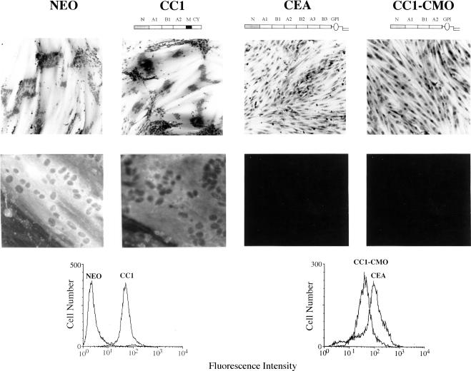
Figure 5.
Effect of the CC1-CMO protein on myogenic differentiation of L6 myoblasts at the morphological and biochemical levels. Top, photomicrographs of hematoxylin-stained cultures of various rat L6 myoblast transfectants incubated in differentiation medium for 7 d. Bottom, photomicrographs obtained by fluorescent microscopy of cultures incubated for 4 d in differentiation medium and stained by anti-myosin monoclonal antibody and fluorescein isothiocyanate-conjugated, anti-mouse IgG. FACS profiles show the relative cell surface expression levels of the indicated proteins. The results were reproducible and were repeated for at least two independently isolated, pooled, total transfectant populations.

