Figure 3.
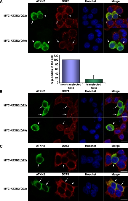
ATXN2 overexpression interferes with P-body assembly. (A) SH-SY5Y cells were transiently transfected with plasmids pCMV-MYC-ATXN2-Q22 or pCMV-MYC-ATXN2-Q79. For staining exogenous ATXN2 and endogenous DDX6, cells were incubated with antibodies directed against the MYC-tag and DDX6, followed by treatment with secondary antibodies coupled to FITC or Cy3, respectively. For quantitative analysis, the percentage of P-bodies in nontransfected versus transfected cells was calculated using the AxioVision software. Here, P-bodies of cells were counted in each picture taken and divided through the cell number. The mean value of P-bodies in the cells counted was calculated and SD was weighted. Then, the number of P-bodies counted for the nontransfected cells was set as 100%, and the number of P-bodies counted for the transfected cells was aligned. (B) Twenty-four hours after transfection, SH-SY5Y cells transiently overexpressing MYC-ATXN2(Q22) or MYC-ATXN2(Q79) or HEK293T transiently overexpressing MYC-ATXN2(Q22) (C) were stained for the MYC-tag and DDX6 or DCP1, respectively. Nuclei were stained with Hoechst. Bars, 10 μm. Arrows indicate transfected cells.

