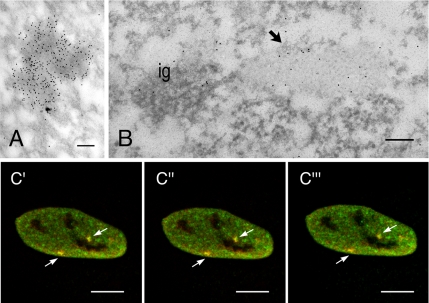
Figure 7.
CF Im68 and PSF colocalize in IGAZs and paraspeckles. (A) Immunolabeling for PSF concentrates on dense fibrillar areas. Bar, 0.1 μm. (B) HeLa cells were double labeled with a rabbit antiserum that recognizes CF Im68 and with mouse mAb that recognizes PSF. PSF (small grains) colocalizes with CFIm (large grains) on IGAZ (arrow) nearby a cluster of interchromatin granules (ig). Bar, 0.1 μm. (C) Fluorescence micrographs of three sequential planes sections through HeLa cells that were double labeled for CF Im68 and for PSF. The images of the green fluorescence of CF Im68 and the red fluorescence of PSF are merged in c′–c‴. Broken arrows indicate colocalization in paraspeckles. Bar, 10 μm.

