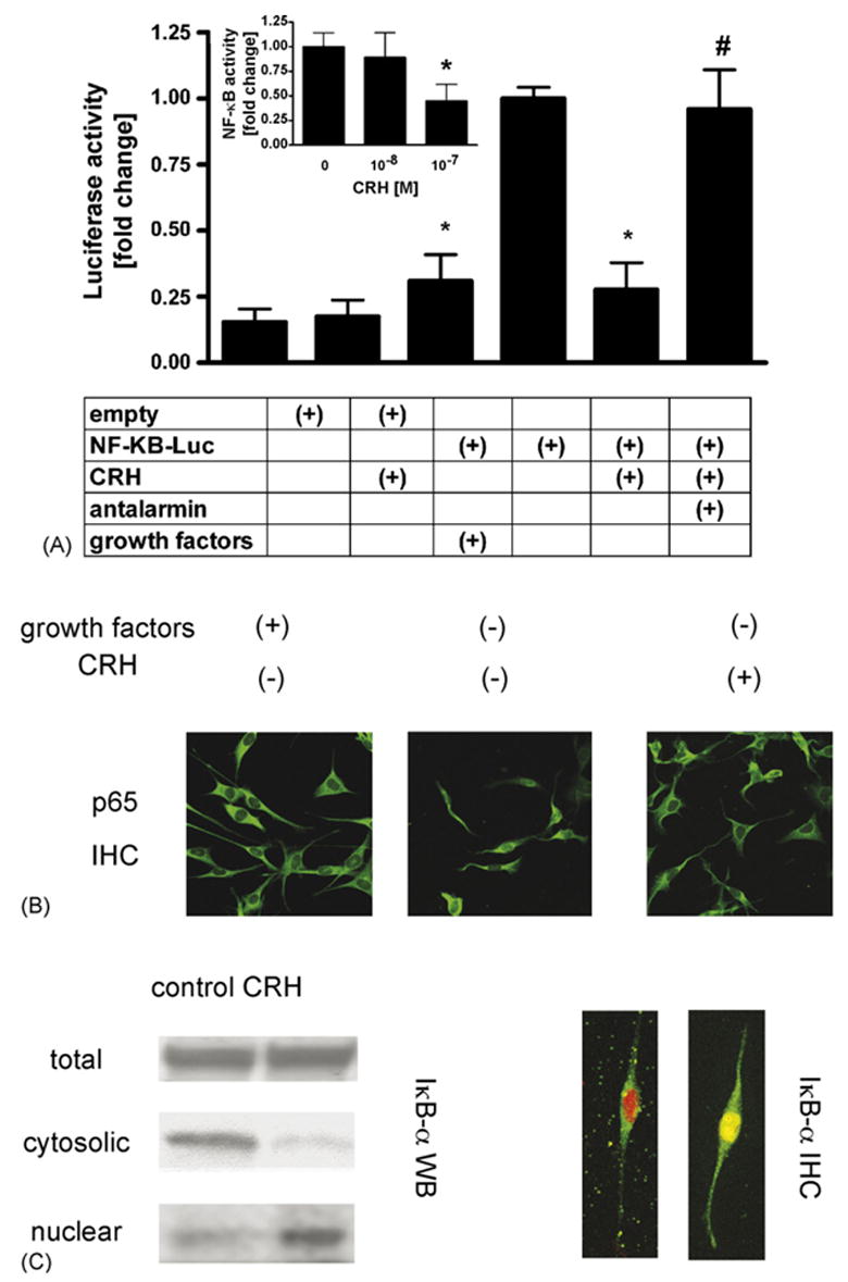
Fig. 1.

CRH decreases NF-κB activity in PIG1 melanocytes and induces translocation of IκB-α from the cytoplasm to the nucleus. (A) Cells were transfected with κB-driven or empty luciferase reporter constructs, pre-incubated in Epilife medium with EDGS (containing growth factors) for 24 h, then medium was changed to Epilife with EDGS or serum free Ham’s F10 and 100 nM CRH was added for 24 h. Antalarmin (10 μM) was added 1 h before medium change. Then cells were lysed and NF-κB transcriptional activity was measured. Inset: effect of increasing doses of CRH on NF-κB activity. Data is presented as mean ± S.E.M. (n = 3; inset: n = 4) in comparison to the cells incubated in medium without growth factors and CRH. *P < 0.05 control vs. CRH. #P < 0.05 CRH vs. CRH and antalarmin. There is no statistical difference between non-treated and treated empty vector controls. (B) Cells were fixed and stained with anti-p65 antibody followed by secondary antibody conjugated to FITC (green). The p65 was distributed in the cytoplasm in the cells incubated in the medium supplemented with serum; incubation of cells in serum free medium resulted in nuclear localization of p65. Incubation of cells with CRH resulted in decreased nuclear localization of p65 and increased cytoplasmic localization. Results are representative of two separate experiments. (C) Cells were pre-incubated in Epilife medium with EDGS for 24 h and then incubated in Ham’s F10 medium without growth factors for 24 h with or without 100 nM CRH. Upper panel: total, cytosolic and nuclear fractions were prepared as described in Section 2. Presence of IκB-α in the lysates separated by SDS-PAGE was estimated with Western blot. Results represent two separate experiments. Lower panel: translocation of IκB-α protein to the nucleus was assessed by confocal microscopy. IκB-α was stained with antibodies conjugated to FITC (green) and propidium iodide was used to stain the nucleus (red). In the absence of CRH IκB-α is localized in the cytoplasm. In the presence of CRH IκB-α was co-localized with nuclear stain (yellow). Result is representative of 4 separate treatments per condition. (For interpretation of the references to color in this figure legend, the reader is referred to the web version of the article.)
