Figure 3.
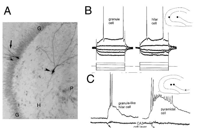
Morphology and electrophysiology of ectopic hilar granule cells born after seizures. A) The morphology of a hilar granule cell from a pilocarpine-treated rat (arrowhead) is shown after intracellular injection of Neurobiotin. For comparison, two granule cells in the granule cell layer (arrow) were also injected. G = granule cell layer; H = hilus; P = pyramidal cell layer. Calibration = 100 μm. B) Physiology of hilar granule cells. Responses to direct current injection (rectangular current pulses, as shown at the bottom) are superimposed for a granule cell located in the granule cell layer (left, “granule cell”) and a granule-like neuron located in the hilus (right, “hilar cell”) of the same slice. This slice was from a pilocarpine-treated rat which had status epilepticus and recurrent seizures. A diagram of the location of these cells is shown at top right. G = granule cell layer, H = hilus, P = pyramidal cell layer. Arrowheads mark spontaneous synaptic potentials. Calibration = 20 mV, 50 msec. C) Comparison of pyramidal cell and ectopic granule cell activity. Simultaneous extracellular recordings from the pyramidal cell layer of CA3b (bottom) and intracellular recordings are shown for a hilar granule cell (left) and a pyramidal cell (right). This slice was from a pilocarpine-treated rat that had status epilepticus and recurrent seizures. A diagram of the location of the cells (dots) and extracellular recording (x) is shown at top right. The arrows point to the population spike of the spontaneous burst discharge recorded extracellularly. Calibration same as B. Used with permission, ref. 87.

