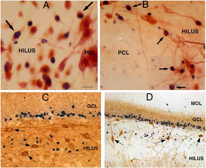
Figure 4.
Ectopic hilar granule cells are born after status epilepticus. A-B: Following injection of BrdU after status epilepticus, tissue sections were double-labeled for BrdU and either NeuN (A) or calbindin (B). The results showed numerous double-labeled neurons (arrows) at the border of the hilus and pyramidal cell layer. Calibration = 30 μm (A) and 60 μm (B). PCL = end of pyramidal cell layer. From ref. 87 with permission: C-D. In a different animal, double-labeling with BrdU and neuropeptide Y (C) or BrdU and parvalbumin (D) demonstrated no double-labeled cells, but numerous hilar neurons that were immunoreactive for neuropeptide Y or parvalbumin (arrows). Calibration (in A) = 60 μm (C) and 120 μm (D).

