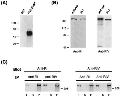
Figure 2.
Western blot analysis of Drosophila CpG MTase-like proteins. (A) Hybridization of GST or NLS-3′hMT polypeptide with anti-RI. The generation of the NLS-3′MT is described in Materials and Methods. The numbers on the left indicate the molecular masses (kDa) of markers. (B) Hybridization of extracts from Drosophila embryos or the Drosophila SL2 cell line. The probes used are anti-RI (Left) and anti-RIV (Right). The approximately 47-kDa band in the “embryo” lane of the right panel is not reproducible and could be the result of degradation. (C) Cross-immunoprecipitation (IP) and Western blot analysis using anti-RI and anti-RIV. Drosophila embryo extract was immunoprecipitated with anti-RI or anti-RIV, and then hybridized with anti-RIV or anti-RI, respectively. Samples on the blots include the total extract (T), supernatants after IP (S), and precipitates from IP (P).

