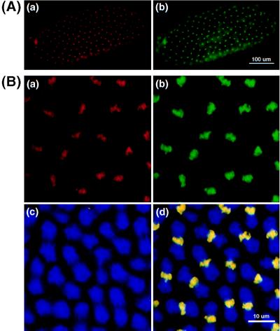
Figure 5.
Colocalization of DmMTR1 with DNA of metaphase chromosomes. (A) Low magnification of metaphase chromosomes of a stage 10 embryo. (B) High magnification of a stage 12 embryo. DmMTR1 (red) is stained with anti-RIV in Aa and Ba. DNA (green) is counterstained with sytox in Ab and Bb. The mitotic α-tubulin (blue) structure is shown in Bc. The superimposed image of DmMTR1, DNA, and α-tubulin is shown in Bd.

