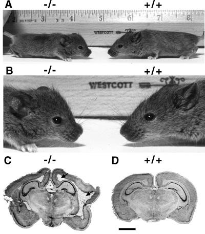
Figure 2.
Hydrocephalus in homozygous Nfia− mice. (A) A homozygous Nfia− pup (−/−) and a wild-type littermate (+/+) are shown. Note the smaller size and foreshortened head of the −/− animal. (B) Enlargement of A showing the characteristic “dome head” of the Nfia− (−/−) animal compared with the more wedge-shaped head of wild-type animal. (C and D) Cresyl violet stained coronal sections through the brains of 6-month-old Nfia−/− (−/−) and wild-type (+/+) animals. Note the dilation of the ventricles (arrows in C). (Bar in D = 2 mm.)

