Figure 3.
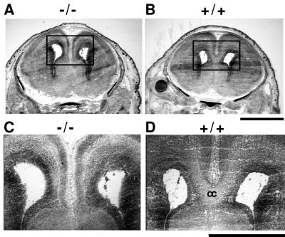
Absence of corpus callosum in Nfia−/− mice. Cresyl violet-stained coronal sections thorough the brains of C57BL/6 fetuses 18 days post coitus. (A and B) Wide-field pictures of the brains of −/− and +/+ littermates with the region surrounding the corpus callosum boxed. (C and D) Expansion of A and B showing the corpus callosum in the +/+ animal (labeled cc in D) and the absence of the corpus callosum in the −/− animal. Serial sections throughout this region showed a complete absence of callosal development in −/− animals. (Bars at the bottom right of B and D equal 2 and 1 mm, respectively.)

