Figure 4.
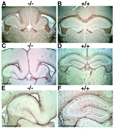
Reduced GFAP expression in Nfia−/− mice. Coronal cryostat sections of the brains of 3-month-old Black Swiss Nfia−/− (−/−) and wild-type mice (+/+) were fixed and stained with antibodies against PLP and GFAP. (A and B) PLP expression in Nfia−/− (A, −/−) and wild-type (B, +/+) mouse brains. Note expanded ventricles indicating hydrocephalus and absence of the corpus callosum (tract lying directly above the hippocampus spanning the two hemispheres) in A vs. B. Expression of PLP appears normal in both animals in the tracts of the fimbria (dark sac-like regions on lower left and right of the hippocampus). (C and D) GFAP expression in Nfia−/− (C, −/−) and wild type (D, +/+) mouse brains. Note the severe reduction in GFAP staining in the cortex and hippocampus (as assessed by reduced levels of punctate cellular brown precipitate), the expanded ventricles, and the absence of corpus callosum in C vs. D. Loss of GFAP staining is regional with reduction of staining in the cortex and hypothalamus but retention of staining with some altered morphology in the fimbria (sacs below the hippocampus) and thalamic nuclei (not shown). (E and F) Higher magnification of GFAP expression in the left dentate gyrus regions of C and D. Note distorted dentate gyrus appearance (probably secondary to hydrocephalus) and reduced GFAP expression in the dentate gyrus in E vs. F. (Bars = 1 mm.)

