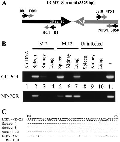
Figure 1.
Detection of LCMV-WE RNA in immune mice. (A) PCR amplification strategy with schematic representation of the S strand of LCMV, which encodes the glycoproteins (GP1 and GP2) and the nucleoprotein (NP) in opposite sense. The primers used are indicated. (B) RT-PCR on reverse-transcribed DNase-treated RNA extracted from spleen, kidney, and lung of two B6 mice (M7 and M12 in Table 3) infected 80 days previously with LCMV-WE (200 pfu i.v. for M7 and 2 × 10⁶ pfu i.v. for M12) using GP- and NP-specific primers as detailed in Material and Methods. Products were run on 1% agarose gels and were visualized with ethidium bromide. Product sizes are 907 bp for GP and 207 bp for NP. Controls in this experiment were no cDNA (lane 1) and RNA extracted from organs of an uninfected mouse (lanes 8–10). The positive control was cDNA template from MC57 cells infected with LCMV (multiplicity of infection 0.02) and was cultured for 48 h. (C) Partial sequence alignment (nucleotides 438–479) from amplified GP products (spleen isolates). Products were sequenced by using inner PCR primers (RC1/DM1) and were compared with LCMV-WE sequence derived from our viral stock (LCMV-WE-ZH) and from a plasmid frequently used in our laboratory, containing GP cDNA (GenBank accession number M22138). Positions of difference in nucleotides are indicated.

