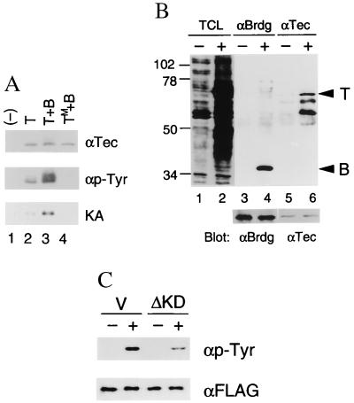
Figure 4.
(A) Activation of Tec by BRDG1. 293 cells were transfected with empty vector (−) or with expression vectors encoding Tec (T), TecKM (TecM), or BRDG1 (B) as indicated at the top. Tec immunoprecipitates prepared from the various transfected cells were then either subjected to immunoblot analysis with antibodies to Tec (αTec) or to phosphotyrosine (αp-Tyr) or assayed for in vitro kinase activity (KA) with [γ-32P]ATP. (B) Ramos cells were left unstimulated (−) or stimulated (+) with anti-human IgM F(ab′)2 fragments (10 μg/ml) for 5 min. Total cell lysates (TCL) (10 μg of protein per lane) and immunoprecipitates prepared with antibodies to either BRDG1 (αBrdg) or Tec (αTec) were then subjected to immunoblot analysis with antibodies to phosphotyrosine (top lane). The positions of Tec (T) and BRDG1 (B) are indicated on the right. The same membrane was reprobed with anti-BRDG1 antibody or anti-Tec antibody as indicated at the bottom. (C) Ramos cells (5 × 10⁶) were electroporated with pcDNA-BRDG-F (10 μg) plus 15 μg of pSRα (V) or pSRα-TecΔKD (ΔKD). After 12 h of culture, cells were treated for 1 h in IMDM/1% FBS at the concentration of 5 × 10⁶/ml and then left unstimulated (−) or stimulated (+) with anti-IgM antibody for 10 min. From each set, FLAG-tagged BRDG1 was immunoprecipitated and probed with either anti-phosphotyrosine antibody or anti-FLAG antibody.

