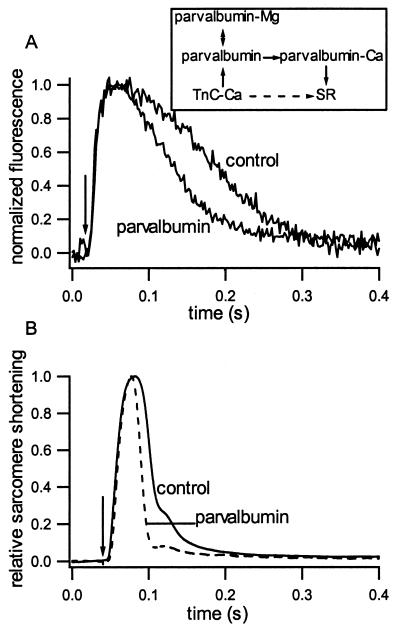
Figure 2.
Fluo-3 fluorescence and mechanical properties in cardiac myocytes. (A) Example of fluorescence intensity vs. time at 37°C. Arrow indicates time of electrical stimulation. (Inset) Schematic of Ca2+ movements during relaxation. Normally, Ca2+ moves from Troponin C (TnC) directly to the SR (dashed arrow). In the presence of parvalbumin, Ca2+ coming off TnC exchanges with Mg2+ bound to parvalbumin where it is temporarily stored before uptake by the SR (solid arrows). (B) Sarcomere movement vs. time as measured by first-order laser diffraction at 37°C from representative control and parvalbumin-expressing myocytes.

