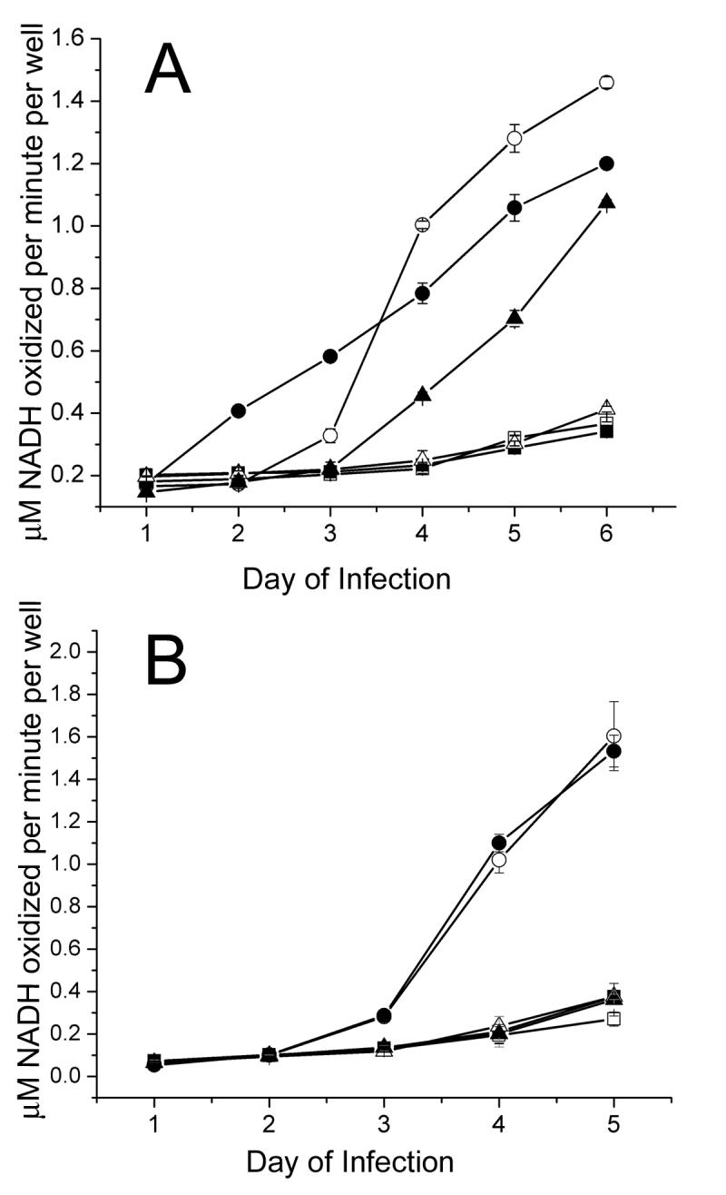
Figure 2.

AAV altered lysis of Ad infected cells. A549 cells were infected with Ad5 or the ADP mutant pm534 in combination with (A) wild-type AAV or (B) a recombinant AAV vector lacking AAV gene expression. Cell lysis results in lactate dehydrogenase (LDH) release into the culture medium, and LDH catalyzes the conversion of NADH to NAD. Therefore increased NADH oxidation, which was measured by the change in absorbance at 340 nm, was indicative of increased cell lysis. Cells infected with Ad5 alone are shown as white circles, and cells coinfected with Ad5 and AAV are shown as black circles. Black and white triangles represent pm534 infections with and without AAV, respectively. Uninfected cells are shown as white squares, and AAV-infected cells are shown as black squares. LDH assays were conducted in triplicate, and the averages are reported (+/− standard deviation). The data shown are representative of three independent experiments.
