
Fig. 1.
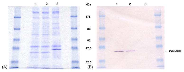
(A) Coomassie blue stained SDS-PAGE of WNV 80E protein expressed by Drosophila S2 cells under non-reducing conditions. Lane 1, Spinner Culture #1 of cell line WN-80E-1 harvested 2/19/03; Lane 2, Spinner Culture #2 of cell line WN-80E-1 harvested 2/10/03; Lane 3, culture of a dengue transformant cell line. The migration of the WNV 80E is faster than the dengue 80E due to differences in glycosylation and tertiary structure (samples are non-reduced). (B) Western blot of duplicate SDS-PAGE gel seen in A. The blot was probed with a commericially available WNV rabbit polyclonal from BioReliance. This antibody cross-reacts slightly with the Dengue 80E.