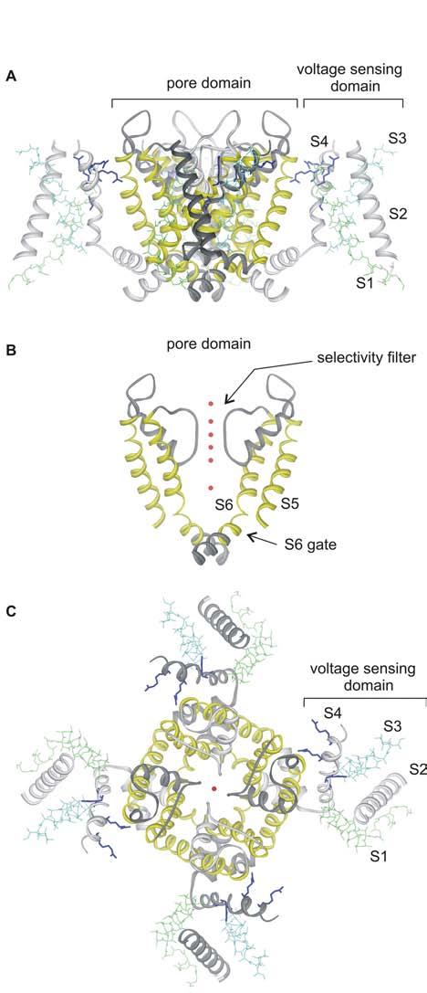
Fig.1.

X-ray structure of the Kv1.2 channel
(A) Side view of the tetrameric Kv1.2 structure oriented so that the extracellular side of the membrane is positioned at the top. (B) Side view with front and back subunits and all four voltage sensing domains removed for clarity. Red spheres are potassium ion bound within the pore, marking the location of the ion permeation pathway. (C) View of the Kv1.2 structure from the extracellular side of the membrane. Images were created us DSViewer Pro and Protein Data Bank accession ID 2A79 (Long et al., 2005).
