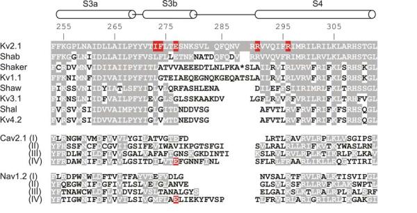
Fig.5.

Sequence alignment for the S3 through S4 paddle region of voltage-activated cation channels.
Horizontal cylinders indicate the approximate positions of S3 (divided into S3a and S3b) and S4 helices. Numbers indicate the residue positions within the Kv2.1 channel and shading indicates similarity to Kv2.1. For the Shaker Kv channel 13 residues have been omitted for alignment purposes and their position marked by the asterisk. Red highlighting indicates residues whose mutations cause pronounced changes in the binding affinity of gating modifier toxins (see text for details).
