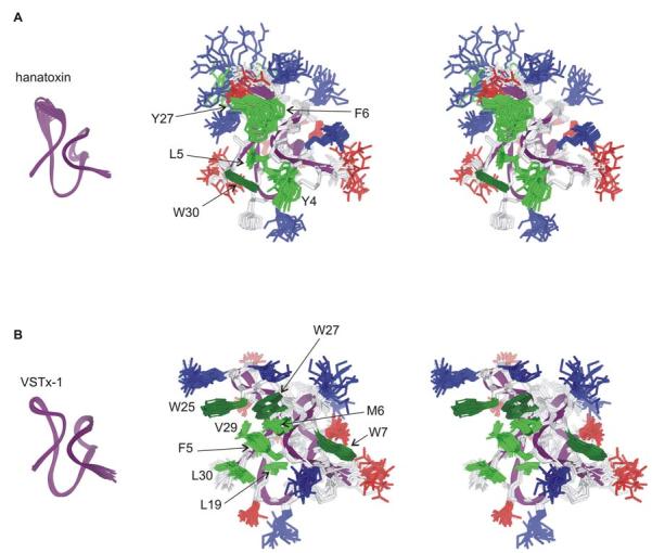
Fig.6.

NMR solution structures of Hantoxin and VSTx-1.
(A) Stereo pairs of the hanatoxin structure represented as a bundle of 20 superimposed conforms. Residue coloring as follows: blue, basic; red, acidic; pink, Ser/Thr; green, hydrophobic (Trp is dark green). Backbone fold is shown at left in purple. (B) Stereo pairs of the VSTx1 structure represented as a bundle of 20 superimposed conforms with coloring as in A. The C-termini in both toxins (F32-S35 in hanatoxin and S32-F34 in VSTx1) are poorly constrained and have been removed for clarity. Images were created us DSViewer Pro and Protein Data Bank accession IDs 1D1H for hanatoxin (Takahashi et al., 2000) and 1S6X for VSTx1 (Jung et al., 2005).
