
Figure 2. Persistent RAD51 Localization in Mre11ATLD1/ATLD1 Mice.
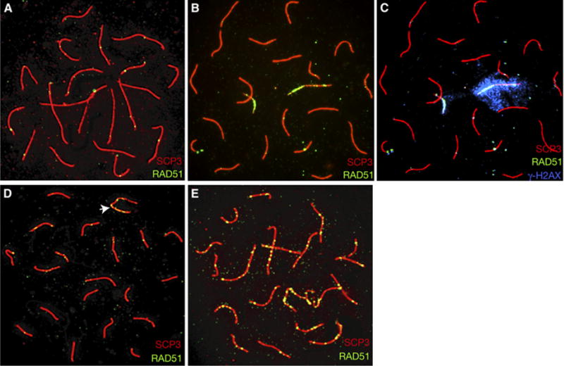
Relative to controls (wild-type oocyte, [A]; wild-type spermatocyte, [D], XY bivalent indicated by arrow), pachytene meiocytes from Mre11ATLD1/ATLD1 mice exhibit prolonged localization of RAD51 foci (mutant oocyte, [B]; mutant spermatocyte, [E]). Persistent RAD51 foci colocalize with γH2AX ([C], same spread as in [B], but overlayed with γH2AX, blue).