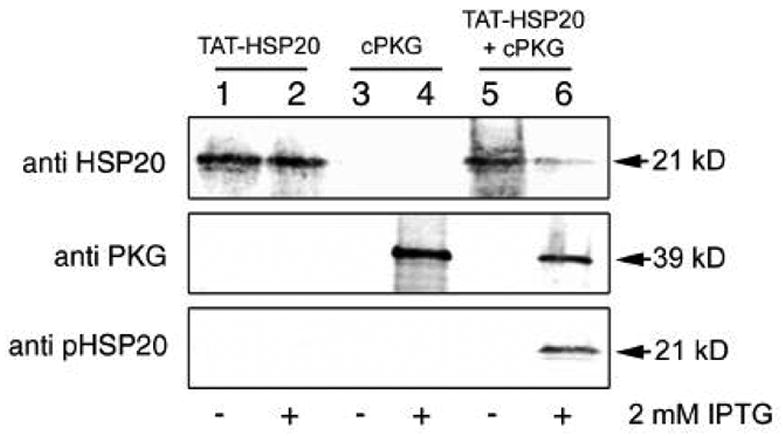
Figure 1.

TAT-pHSP20 is expressed and phosphorylated in E. coli. Representative immunoblot analyses of BL21(DE3) bacterial extracts taken from a large scale (4L) co-expression experiment. Extracts were generated before (-) or after (+) induction with 2 mM IPTG and 20 μg protein was loaded per well. Lanes 1 and 2 - pET14b TAT-HSP20 vector only; lanes 3 and 4 - pACYC Duet PKG C subunit vector only; lanes 5 and 6 - pET14b TAT-HSP20 and pACYC Duet PKG C subunit vectors together. Blots were probed with the antibodies indicated on the left.
