
Fig. 1. UVC and IR activate different signaling intermediates in human cells.
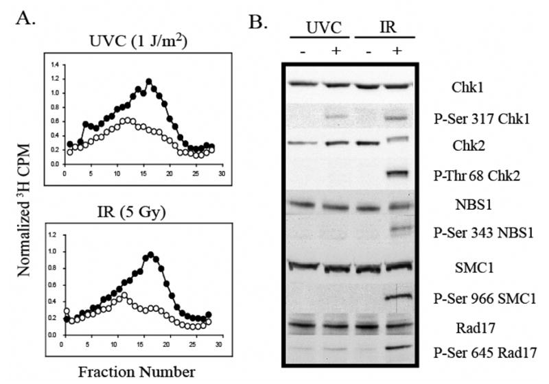
A.) NHF1 cells were grown in the presence of [14C]thymidine for ~ 40 h to label DNA uniformly, and then in non-radioactive medium overnight. Cells were sham-treated or exposed to UVC (1 J/m2) or IR (5 Gy), incubated at 37 °C for 30 min, and then labeled for 15 min in medium containing [3H]thymidine. Cells were harvested and nascent DNA separated by velocity sedimentation. Net 3H radioactivity corrected for 14C spillover was normalized to cell number (total 14C radioactivity). Closed circles (●) represent profiles from sham-treated cells while grey circles (○) represent those from irradiated cultures. B.) Normal human fibroblasts were sham treated or irradiated with either 1 J/m2 UVC or 5 Gy IR. Cells were harvested 1 h after irradiation and cell extracts prepared for western immunoblot analysis.