
Fig. 6. Over-expression of Dbf4 reverses the UVC-induced S checkpoint.
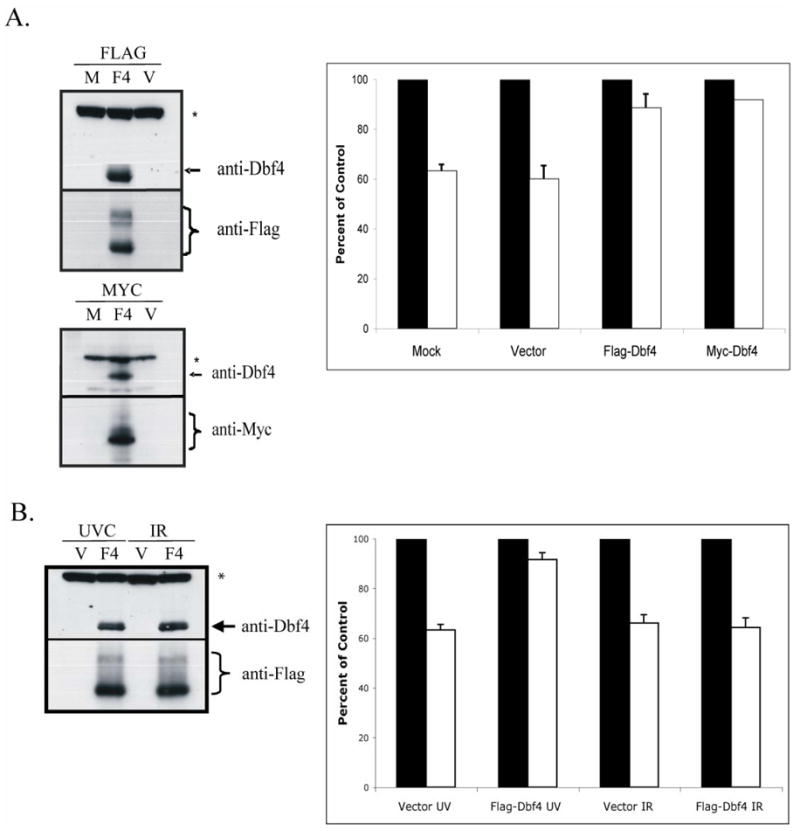
A.) HeLa cells were mock-transfected (M), transfected with an empty vector (V), or transfected with Flag-, or Myc-tagged Dbf4 expression vectors (F4). Cells were harvested 48 h later and extracts prepared for western immunoblot analysis (* = non-specific band). In separate experiments, transfected cultures were incubated with [14C]thymidine to label DNA uniformly; then, cells were treated with either 0 or 1 J/m2 UVC, incubated for 30 min at 37°C and pulsed-labeled with [3H]thymidine for 15 min. DNA synthesis was measured as described in the legend to Fig. 2C and graphed as percentages of paired, sham-treated controls [n=5 (Mock), n=7 (Vector), n=5 (Flag), n=2 (Myc); black bar, sham-treated controls; white bar, UVC-irradiated cells. Error bars correspond to one standard deviation of the mean]. B.) HeLa cells were transiently transfected with either empty vector (V) or a Flag-Dbf4 expressing vector (F4). Cells were harvested 48 h later and extracts prepared for western immunoblot analysis (* = non-specific band). In separate experiments, transfected cultures were incubated with [14C]thymidine to label DNA uniformly; then, cells were sham-treated or irradiated with either 1 J/m2 UVC or 5 Gy IR, incubated for 30 min at 37°C and pulse-labeled with [3H]thymidine for 15 min. DNA synthesis was measured as described in the legend to Fig. 2C and graphed as a percentages of paired, sham-treated controls (n=3; black bar, sham-treated controls; white bar, UVC- or IR-treated cells. Error bars correspond to one standard deviation of the mean).