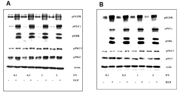
Figure 1.

Immunoblotting data for EGF treatment of 5 minutes (A) and 1 hour (B) across different fibronectin concentration of surfaces. Tissue culture plates were coated with different fibronectin (FN) concentrations. NR6WT cells were grown on these surfaces for 24 hours in complete growth medium and quiesced for another 24 hours in medium containing 0.5% dialyzed FBS. EGF was added for a period of 1 hour, cells washed once with PBS and lysed. Cell lysates were resolved using SDS-PAGE and immunoblotted using specific antibodies for various phosphorylated proteins. At least 5 replicates for each signaling protein were created for polynomial modeling. Actin served as a loading control.
