Figure 4.

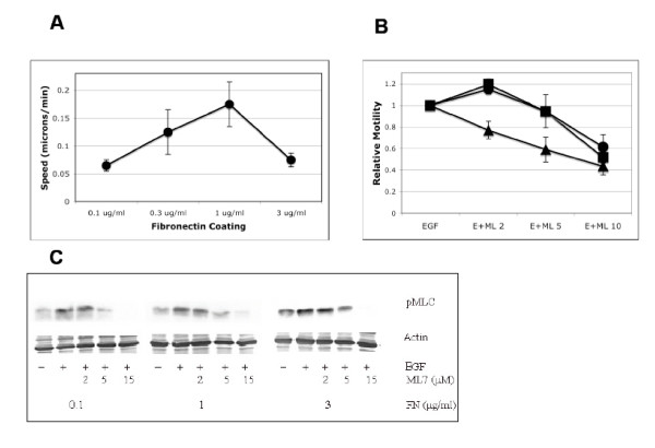
Subtotal inhibition of myosin light chain kinase increase cell migration via single-cell tracking. NR6WT fibroblasts were grown on fibronectin-coated surfaces coated and quiesced in serum-restricted conditions for 16 hours. After drug inhibition and/or EGF stimulation, single cells were tracked for up to 20 hours and their migration speeds analyzed using Visible, developed by Reify Corporation. Each experimental condition is the average ± SEM of 15–20 cells. (A) Four concentrations of fibronectin were used (0.1, 0.3, 1, 3 μg/ml) and the biphasic relationship between speed and fibronectin was indeed reproduced via our single-tracking setup and analysis. (B) Under higher fibronectin conditions (1 and 3 μg/ml), partially inhibitory ML-7 concentrations increases migration speed while further inhibition reduces the closure of the in vitro wound. At low fibronection concentrations (0.1 μg/ml) further reduction of MLC activation reduced wound closure. Shown are mean ± SEM of four experiments performed in triplicate and normalized within run to no ML-7 control speeds. In comparison to no ML-7 treatment, P < 0.05 for 2 μM ML-7 treatments on 1 and 3 μg/ml fibronectin; the decreases in speed were also statistically significant at higher ML-7 concentrations for all three surfaces. 0.1 μg/ml FN are triangles, 1 μg/ml FN are circles and 3 μg/ml FN are squares. (C) Attenuation of MLC activity using graded concentrations of MLCK inhibitor, ML-7. MLC activity is completely abrogated at concentrations greater than 15 μM. Three FN levels (low, medium and high concentrations) are shown for simplicity. Shown is one of three representative experiments.
