
Fig. 1.
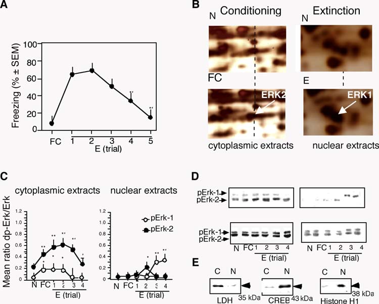
Up-regulation of hippocampal Erk-1 and Erk-2 during fear conditioning and extinction. (A) Time course of fear extinction revealed by decrease of freezing after nonreinforced contextual exposures. (B) Representative proteomic scans of cytoplasmic extracts obtained 1 hr after training (left), demonstrating increased phosphorylation of pErk-2; and nuclear extracts obtained 1 hr after the 4th extinction trial (right) demonstrating nuclear accumulation of Erk-1. (C) The levels of cytoplasmic pErk-1 and pErk-increased significantly after conditioning and extinction (left panel). Nuclear pErk-1 increased significantly after the 3th and 4th extinction trial (right panel). (D) Representative immunoblots. (E) The quality of nuclear extracts was examined by immunoblot experiments with antibodies detecting the nuclear proteins CREB and histone H1 and cytoplasmic protein LDH. Enrichment of the nucleoproteins was observed only in the nuclear extracts. The number of mice per group was 5. Statistically significant differences obtained by one-way ANOVA followed by the Scheffés test for multiple comparisons: *p < 0.01 vs N group; **p < 0.001 vs N group.