
Fig. 2.
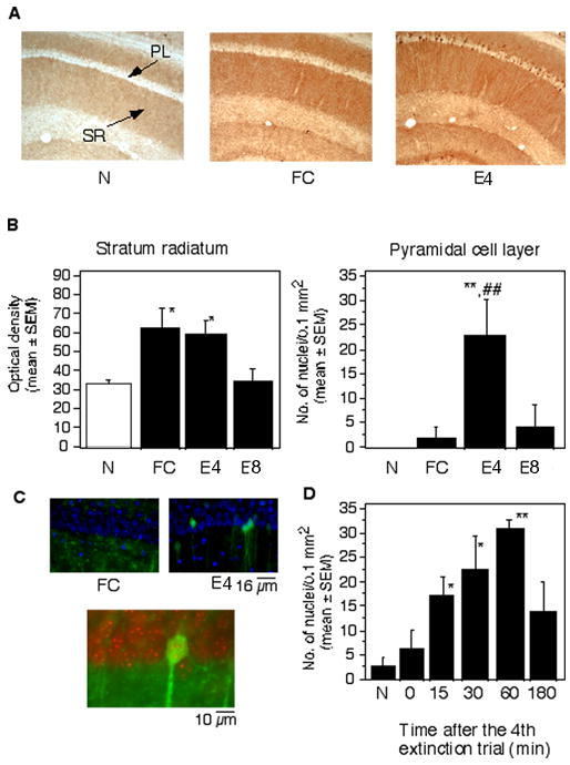
Localization of pErk-1/2 after conditioning and extinction. (A) Representative photomicrographs demonstrating the level of pErk-1/2 in the CA1 hippocampal area of mice of the N, FC and E groups. (B) Mean density of pErk-1/2 immunoreactivity in the stratum radiatum of the experimental groups (left). Mean number of cells per CA1 hippocampal area of the experimental groups (right). Statistically significant differences obtained by one-way ANOVA followed by the Scheffés test for multiple comparisons: *p < 0.01 vs N; **p < 0.001 vs N and T. PL, pyramidal cell layer; SR, Stratum radiatum. (C) Nuclear pErk-1/2 was not observed in mice exposed to training (upper left), whereas extinction triggered a significant increase of nuclear pErk-1/2 in hippocampal pyramidal neurons (upper right), as revealed by merged images with DAPI (blue) and pErk-1/2 (green). Below: DAPI digitized red and pErk-1/2 green show yellow overlapping signals. The mean percentage of Erk-1/2 positive nuclei in the extinction and training groups was 15.4 ± 7.8 and 0.5 ± 1.2, respectively. The number represents a mean ratio of Erk-/2 positive of a total of 200 nuclei counted in a single focal plane in 5 randomly chosen sections of the dorsal hippocampus/mouse. (D) The time course of pErk-1/2 up-regulation after the 4th extinction trial revealed a significant activation already after 15 min whereas 3 hr later the activated kinase was not detectable. Statistically significant differences were revealed by one-way ANOVA followed by Scheffés test : *p < 0.01, **p < 0.001 vs group sacrificed immediately after the extinction trial (0 min). The number of mice per group was 6 for each experiment.