
Figure 3. Expression of lineage-specific cell surface markers on Myc5 cells under different culture conditions.
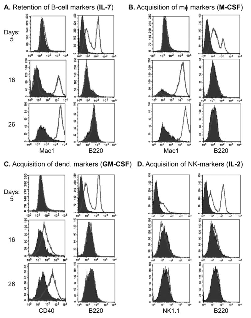
Panels A-D represent growth media supplemented with cytokines favoring particular lineages: B-cell (interleukin-7, or IL-7), macrophage (macrophage colony-stimulating factor, or M-CSF), dendritic (granulocyte-macrophage colony-stimulating factor, or GM-CSF), and natural killer (NK) cell (interleukin-7, or IL-2). B220, Mac-1, CD40, and NK1.1 are the corresponding surface markers. All cultures were assayed on days 5, 16, and 26 following cytokine stimulation using flow cytometry. Filled and open plots represent unstained and antibody-stained cells, respectively. For more details, see Methods and Materials.