
Figure 5. Phagocytosis by Myc5 cells.
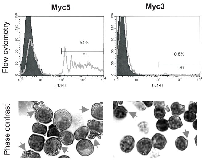
Myc5 and control Myc3 cells were grown in media supplemented with M-CSF. Cells pre-incubated with fluorescent latex beads were subjected to flow cytometry (top panels) and phase-contrast microscopy (bottom panels). In the top panels, filled and open plots refer to cells without and with beads, respectively. In the bottom panels, arrows point at cells with ingested or attached beads. For more details, see Materials and Methods.