
Figure 6. Lymphoid- and macrophage- specific gene expression is affected by Pax5.
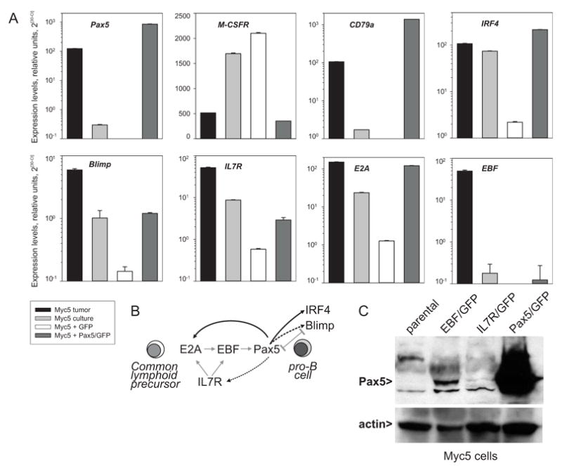
A Real-time RT-PCR analysis of Pax5, M-CSFR, CD79a, IRF4, Blimp, IL7R, E2A, and EBF transcript levels in the Myc5 tumor, cultured Myc5 cells, and cultured Myc5 cells transduced with the empty MIGR1 vector (“GFP”) or the Pax5/MIGR1 virus (“Pax5/GFP”). Gamma-actin was used as an internal control. Relative expression levels were calculated as described in Methods and Materials. B. Interactions between IL7R and B-lymphoid transcription factors during differentiation of common lymphoid progenitor (CLP) towards the pro-B-cell. Grey arrows indicate known interactions (modified from [1]). Black arrows refer to data in A. Dotted arrows indicate partial restoration of mRNA levels by retrovirally encoded Pax5. C. Immunoblotting with anti-Pax5 and anti-actin antibodies. Cells used were parental Myc5 cells and their EBF-, IL7R-, and Pax5-transduced derivatives.