
Figure 2.
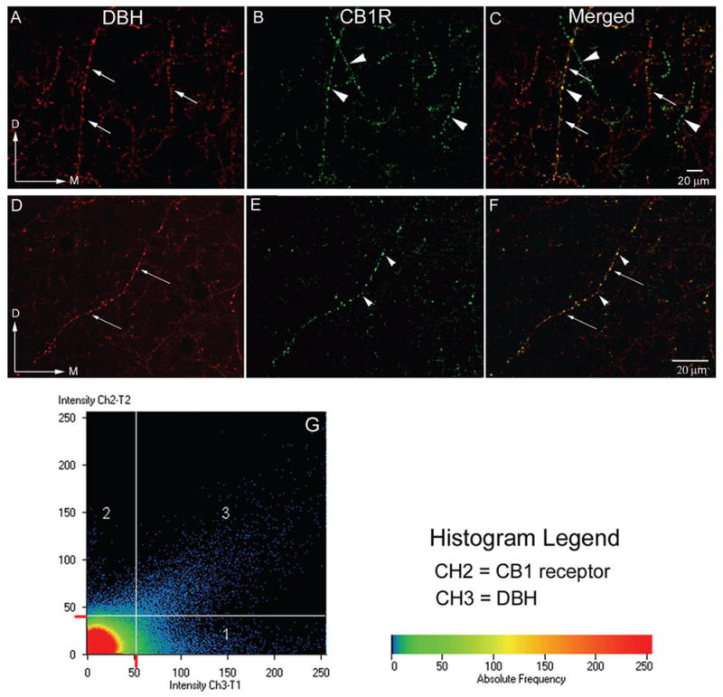
A–F: Confocal microscopy photomicrographs showing sections processed for the immunocytochemical detection of the catecholamine synthesizing enzyme, DßH (panel A, D) and CB1 receptors (panel B, E) in coronal sections from the infralimbic area of the frontal cortex. Abundant labeling in varicose processes was present in the frontal cortex for both antigens. Processes containing DßH alone can be seen at the long white arrows while fibers containing CB1 alone are shown at arrowheads. Panels C and F are merged images. Yellow labeling denotes co-localization of both CB1 and DßH in the same process. Magnification A–C: 20x, D–F: 40x oil G. Histogram analysis demonstrating the co-localization of DßH (red) and CB1 receptor (green). Intensity of labeling for DßH is seen on the x-axis (CH2-T1) and intensity of labeling for the CB1 receptor is on the y-axis (CH3-T2). Colors in the histogram represent pixel frequency and intensity.