
Figure 3.
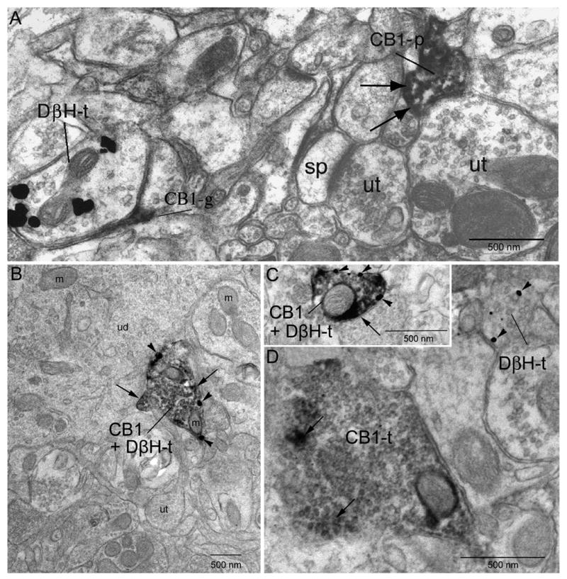
Electron micrographs showing localization of CB1 and DßH in axon terminals (t) in the frontal cortex. A. Dense peroxidase labeling for CB1 can be identified in a cellular process (CB1-p) denoted at black arrows. The CB1-p is apposed to an unlabeled terminal (ut). Another ut nearby contacts a spine (sp). The DßH-t is apposed by a profile resembling a glial process that is immunoreactive for CB1. B. Co-localization of gold-silver labeling for DßH (arrowheads) and peroxidase labeling (indicated by long black arrows) for CB1 in the same axon terminal. The DßH + CB1 terminal is apposed to an unlabeled dendrite (ud). Several unlabeled terminals can be seen in the neuropil. C. Reversal of immunolabels indicates similar patterns of immunolabeling. Gold-silver labeling for the CB1 receptor (arrowheads) and peroxidase labeling for DßH (long black arrows) can be seen in the same terminal. Gold-silver labeling for the CB1 receptor shows localization (CB1-t) to the plasma membrane of the axon terminal. D. A CB1 receptor immunoperoxidase-labeled terminal and a DßH immunogold-labeled terminal converge (DßH-t) onto a common dendrite. m: mitochondria. ut: unlabeled terminal. sp: spine.