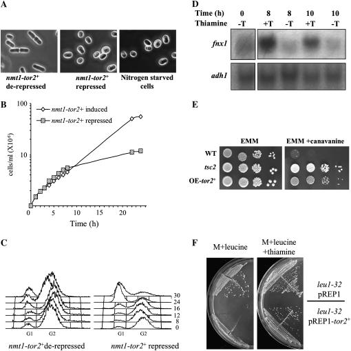
Figure 1.—
tor2 phenotypes. (A) Left and middle: tor2− cells expressing nmt1-tor2+ (TA313) were grown to midlog phase and transferred to fresh minimal medium in the absence (derepressed) or presence (repressed) of thiamine. After 12 hr, cells were visualized by light microscopy. Right: wild-type cells (TA001) were starved in minimal medium containing no nitrogen for 21 hr. (B) Δtor2 mutant cells (TA313) were grown as described above. At time zero thiamine was added and cells were counted. (C) FACS analysis of the cells sampled in B. (D) Northern blot analysis. Total RNA was prepared from samples taken at the indicated time points (hours). Northern blots were probed with fnx1+ and with adh1+ as a loading control. (E) Wild-type (TA001), Δtsc2 (TA450), and Δtor2 carrying nmt1-tor2+ (TA313) cells were grown in EMM to midlog phase. Four different dilutions were spotted on EMM plates with and without 60 μg/ml canavanine. Plates were incubated at 30° for 3 days. (F) Cells auxotrophic for leucine were transformed with vector only or with vector containing the nmt1-tor2+ construct. Slow-growth phenotype is observed only when the nmt1 promoter is derepressed (no thiamine). Cells were streaked on minimal (M) plates supplemented with 75 μg/ml leucine and incubated at 30° for 4 days.

