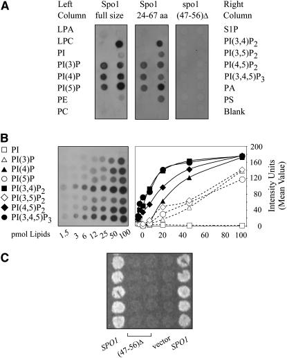
Figure 7.—
Binding of the Spo1 protein to various lipids. (A) Lipid binding using PIP strips with test lipids indicated in corresponding positions on the left and right sides of the strip. LPA, lysophophatidic acid; LPC, lysophosphocholine; PI, phosphatidylinositol; PI(3)P, phosphatidylinositol-3-monophosphate; PI(4)P, phosphatidylinositol-4-monophosphate; PI(5)P, phosphatidylinositol-5-monophosphate; PE, phosphatidylethanolamine; PC, phosphatidylcholine; S1P, sphingosine-1-phosphate; PI(3,4)P2, phosphatidylinositol-3,4-biphosphate; PI(3,5)P2, phosphatidylinositol-3,5-biphosphate; PI(4,5)P2, phosphatidylinositol-4,5-biphosphate; PI(3,4,5)P3, phosphatidylinositol-3,4,5-triphosphate; PA, phosphatidic acid; PS, phosphatidylserine; the bottom right spot does not contain any lipids. Binding is shown for full-size Spo1 (left), a 5-kDa N-terminal fragment (residues 24–67, center), and a derivative of full-size Spo1 with the lysine stretch deleted (residues 45–56, right). (B) Typical results of lipid-binding assays and their quantification for the full-size Spo1 protein using PIP arrays. Test PI and PIP lipids (abbreviated as above) are shown at the left. (C) spo1Δ complementation by wild-type SPO1, its deletion derivative with an excised lysine stretch motif, and no insert in a high-copy plasmid. Dityrosine spore fluorescence is shown for strains sporulated at 23° for 5 days.

