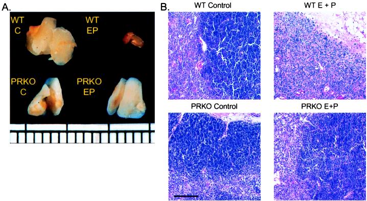
Figure 1.
Gross morphology and H&E histology of E + P treated wild-type and PRKO thymuses. (A) Photographs of wild-type and PRKO thymuses after 12 days of E + P treatment demonstrating lack of involution in PRKO mice. Ruler demarcated in millimeters. (B) Five micrometers H&E-stained sections of thymuses taken from wild-type and PRKO mice treated for 12 days with E, E + P, or vehicle alone. Notice the complete loss of cortical lymphocytes in the wild-type E + P-treated mouse, whereas the PRKO thymus appears intact. Bar = 100 μm.

