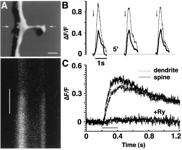
Figure 1.
Transient calcium responses of dendrites and spines to caffeine. (A) A sample illustration of a dendrite/spine segment loaded with Oregon green-1 and 3D-reconstructed. Arrows indicate the location of the line scanned across the spine/dendrite axis, before and during application of caffeine. (Bar = 1 μm.) (Lower) A line scan as described above. Each line is scanned for ≈0.8 msec, and the scan seen comprises ≈400 msec, from top to bottom. Caffeine (left vertical bar, 150 msec) caused a rise in calcium-sensitive fluorescence, with about the same latency in the spine and parent dendrite. (B) Example of repeated responses to caffeine in a single spine/dendrite segment comprising a spine (thin line) and dendrite (thick line). Caffeine is applied once every 5 min (arrows) with no significant reduction in magnitude of the response. (C) Summary of spine/dendrite pairs comparing responses to caffeine in control (n = 152 spines) and the presence of ryanodine (Ry, n = 41 spines). The half-time for recovery from the caffeine action is ≈1.2 sec.

