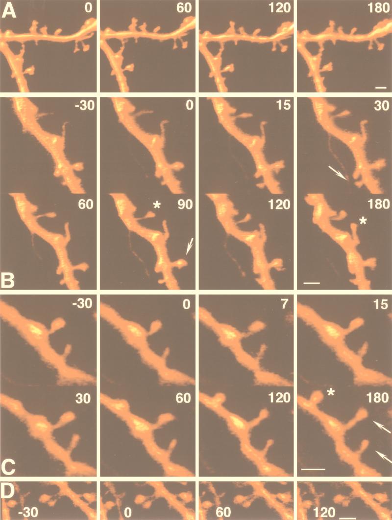
Figure 2.
Morphological changes in spines after exposure to caffeine. Cells were stained with calcein and were 3D-reconstructed in the confocal microscope at the times indicated in minutes. (A) Control dendritic segment, imaged at 1-hr intervals, showing no difference in shape or length of the dendritic spines. (B and C). Two dendritic segments, imaged before and after exposure to caffeine, applied around time 0. Note the formation of a spine head (arrows), the elongation of the spine neck of the bottom spine, and the change in position in space of the spine (asterisk). (C) Like B, a dendritic segment is exposed to caffeine at time 0, and a novel spine appears at 120 min after the onset of the caffeine treatment (asterisk). (D) Exposure to caffeine in the presence of ryanodine, under the same conditions seen in B and C. No change in spine shape or length were seen in this experiment. (Bar = 2 μm.)

