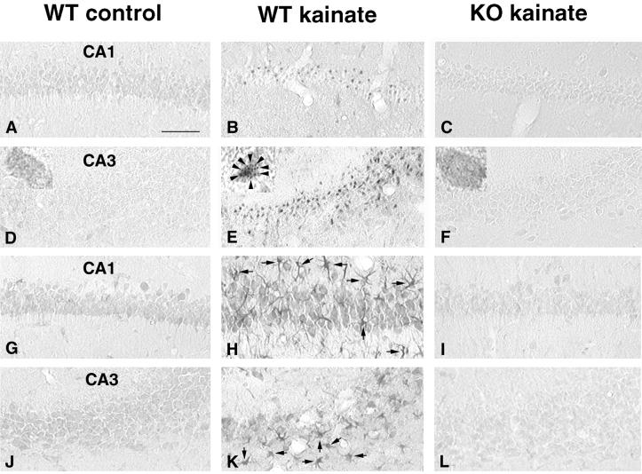
Figure 4.
Kainate-induced expression of bax and caspase 3 in PPT-A(+/+) and PPT-A(−/−) mice. (A-F) Immunocytochemical analysis of bax. In saline-treated controls (A and D), there were few bax-ir neurons, and bax-ir was distributed throughout the cytosol (Inset in D, ×100). Expression of bax was markedly increased in the CA1 (B) and CA3 (E) regions in kainate-treated PPT-A(+/+) mice. Bax-ir was redistributed in punctate fashion (arrowhead in Inset in E, ×100) in PPT-A(+/+) mice 3 days after injection of kainate (35 mg/kg). In contrast, no increase in bax-ir was found in hippocampi (C and F), and bax-ir was not redistributed (Inset in F, ×100) in PPT-A(−/−) mice treated with the same dose of kainate. (G–L) Immunocytochemical analysis of caspase 3-ir. In the saline-injected mice, a small amount of caspase 3-ir was detected in hippocampus (G and J). Three days after injection of kainate (35 mg/kg), there was a dramatic increase in caspase 3-ir in neurons through the entire pyramidal layer of PPT-A(+/+) mice (H and K); there was also a dramatic increase in caspase 3-ir in glial cells over the entire hippocampus (arrows in H and K). By contrast, there was only minimal induction of caspase 3-ir in CA1 (I) and CA3 (L) in PPT-A(−/−) mice. (Scale bar: 125 μm).

