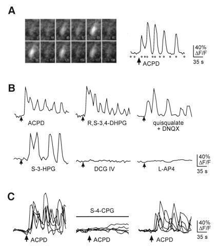
Figure 1.
Activation of intracellular calcium ([Ca2+]i) oscillations by group I mGluRs in developing neocortex. (A) Example of an oscillatory intracellular free calcium response caused by the mGluR agonist trans-ACPD (40 μM). (Left) The fluorescence of a single fluo-3-loaded neuron at several times during imaging. The elapsed time is shown in the lower right corner of each frame. (Right) This cell’s response, measured as % ΔF/F. Open circles indicate the times of the corresponding frames shown at left. ACPD application began at the arrow and continued until the end of the imaging series. (B) The group I mGluR agonists R,S-3,4-DHPG (100 μM), quisqualate (10 μM) in the presence of DNQX (10 μM), and S-3-HPG (300 μM) all produced oscillatory [Ca2+]i responses, whereas agonists of group II mGluRs (DCG-IV, 1 μM) and group III mGluRs (l-AP4, 100 μM) did not produce [Ca2+]i oscillations in these cells. (C) Oscillatory [Ca2+]i changes induced by trans-ACPD were reversibly abolished by the specific group I mGluR antagonist S-4-CPG (1 mM). Five oscillatory responses to trans-ACPD are shown in the Left, and the block of this response and subsequent recovery in the same neurons are shown in the following traces.

