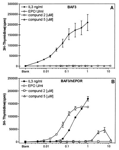
Figure 5.
Mitogenic responses in BAF3 and BAF3/hEPOR cells. BAF3 or BAF3/hEPOR cells were treated with different amounts of EPO, IL-3, or compounds for 48 hr. 3H-thymidine incorporation, as an indicator of cell proliferation, was measured after addition of 4 μCi/ml of 3H-thymidine during the last 4 hr of incubation, as described in Materials and Methods. (A) Amount of 3H-thymidine incorporated in BAF3 cells after different treatments. (B) Effect of same in BAF3 cells expressing human EPOR. Data represent mean (+/− SEM) of two to three experiments where each determination was made in triplicate. Value of x axis reflects the concentration of EPO, IL-3, or compounds in units/ml, ng/ml, and μM, respectively. All assays were performed in 1% DMSO. Values shown above blank on graphs refer to level of radioactivity incorporated in the untreated cultures in the presence of 1% DMSO.

