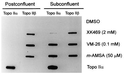
Figure 5.
Covalent binding of topoisomerase IIα and topoisomerase IIβ to cellular DNA after exposure of cells to topoisomerase poisons. Subconfluent and postconfluent human breast cancer cells (MCF-7) were treated with S(−)XK469, VM-26, m-AMSA, or the solvent, DMSO, for 15 min and then were lysed with GuHCl as described (see Materials and Methods). The cellular DNA was banded by CsCl gradient ultracentrifugation, and the CsCl then was removed by dialysis of the pooled DNA fractions. A 30-μg DNA aliquot was removed, and MgCl2 was added to a final concentration of 5 mM. The DNA was digested with protease-free DNase I (Boehringer Mannheim, 0.1 units/ml, 37°C, 1 hr). The aliquot then was applied to a poly(vinylidene difluoride) membrane with a slot blot device. Purified human topoisomerase IIα (TopoGen) also was applied as a control. Blotted proteins were probed with antibodies to human topoisomerase IIα and human topoisomerase IIβ (indicated at the top of each column).

