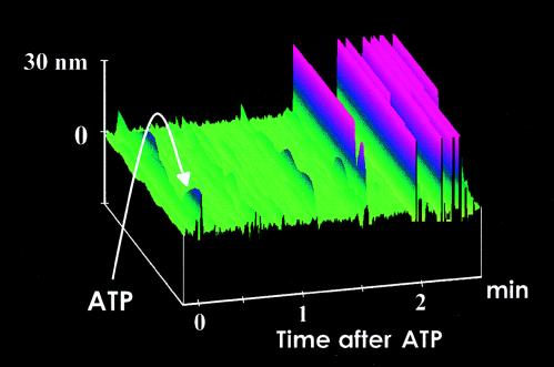
Figure 2.
Functionalized S1 tip scanning the surface of mica in fluid. After the addition of ATP (10 nM) indicated by the arrow, the tip starts to deflect after a time lag of 1–2 min because of diffusion of ATP toward the AFM tip. Tip deflections are shown in pink, whereas the mica surface is shown in green.

