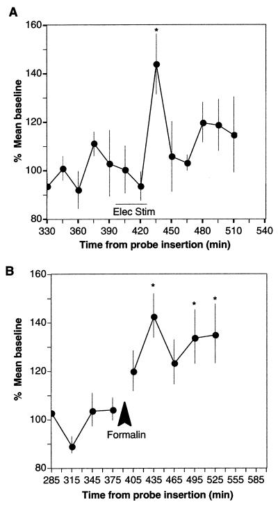
Figure 4.
The release of anandamide in the PAG of the rat stimulated by electrical depolarization or pain. (A) Increased extracellular levels of anandamide after electrical stimulation of the PAG in urethane-anesthetized rats. After the establishment of stable baseline values, electrical stimulation (bipolar 0.1 msec/1 mA, 60-Hz, 5-sec trains with 5-sec rest intervals) was delivered for 30 min. Microdialysis samples were collected in 15-min intervals and analyzed by HPLC with detection by APCI–MS, with selected ion monitoring mode at molecular weight 348.3 (n = 5, P < 0.05, in repeated measures analysis of variance). The asterisks mark points that are significantly different from the baseline average by posthoc test (P < 0.05). The delay in measurement presumably reflects the time needed to produce anandamide in the extracellular space with sufficient overflow to achieve recovery by microdialysis. (B) Increased extracellular levels of anandamide in the PAG after induction of prolonged pain in urethane-anesthetized rats. After the establishment of a stable baseline, a 4% 150-μl formalin solution was injected subcutaneously in both hindpaws. The samples shown span 30-min intervals (n = 6; P < 0.001, repeated measures analysis of variance).

