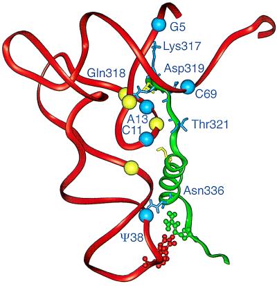
Figure 6.
Molecular structure of part of the tRNAGln–GlnRS complex. The diagram shows the Thr-316–Arg-341 motif of GlnRS and the phosphate-sugar backbone of tRNAGln based on the 2.5-A resolution structure of the complex (6). Amino acid side chains shown are Thr-316, Lys-317, Gln-318, Asp-319, Thr-321, Ser-326, Asn-336, and Arg-341. Spheres representing the phosphate-sugar backbone of residues that interact with the motif are shown (P of residues 5, 8, 11, 12, 13, 14, 25, 38, and 69). Blue spheres indicate nucleotides that are mutationally identified in this work; they interact with amino acid side chains indicated in blue. Arg-341 interacts with the base of U35. Table 2 lists the interacting atoms. The figure was made with insight ii (96.0.6).

