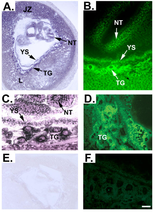
Figure 1.

In situ hybridization and immunolocalization of RFC during early murine embryonic development. Panels A and C demonstrate expression of RFC mRNA in a section of uterus containing an E10.0 embryo. Panel E demonstrates the absence of background staining when a sense riboprobe for RFC is hybridized with placental sections as a negative control. To determine whether RFC protein may be localized to the same areas that are positive for RFC message during murine embryonic development, immunofluorescence was utilized in E10.0 frozen uterus sections. Panels B and D indeed verify that RFC protein is localized in a manner consistent with the expression pattern of RFC message. Specificity of the RFC antibody is demonstrated by lack of fluorescence in tissues incubated with RFC antibody that had been pre-incubated with blocking peptide (Panel F). L, labyrinth zone; NT, neural tube; JZ, junctional zone; TG, trophoblastic giant cells; YS, yolk sac. Scale bar represents 25 μm in A and E and 4 μm in B, C and D.
