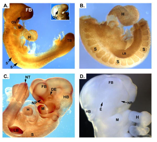
Figure 3.
Immunohistochemical detection of RFC in developing mouse embryos. Panels A and B demonstrate staining for RFC in an E9.0 and E9.5 embryo, respectively. Positive signal is detected throughout the embryo, with the signal being more intense in the optic vesicles, mandible, heart, somites, limb buds and tail bud. At E10.5 (panel C) expression of RFC is present in the aforementioned tissues as well as in the neural tube, nasal pits and developing eye. Control embryos incubated with pre-blocked antibody demonstrated no signal (Panel D). optic vesicle, OV; mandible, M; heart, H; somites, S; limb buds, LB; tail bud, TB; neural tube, NT; nasal pits, NP; developing eye, DE. Scale bar represents 50 μm in A, C, E and F and 100 μm in B and D.

