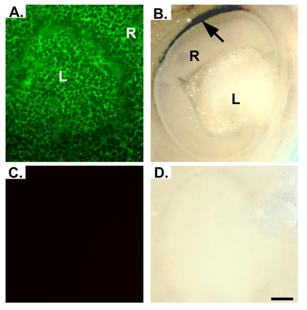
Figure 4.
Immunofluorescent and immunohistochemical detection of RFC in developing mouse eye. Panel A demonstrates immunofluorescent signal for RFC present throughout the developing retina and lens of an E10.0 ICR mouse embryo. As development continues through E12.5, signal in the lens diminishes, while the signal in the retina becomes confined to the RPE (Panel B). Expression appears to be greater in the dorsal region of the retina (black arrow). Sections incubated with pre-blocked antibody demonstrated no signal (Panels C and D). R, retina; L, lens. Scale bar represents 10 μm.

