Figure 6.
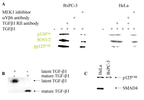
Enhanced level of cytoskeletal anchored proteins in response to TGFβ1 (A). Western Blot analysis of BxPC-3 and HeLa cells as indicated after stimulation with TGFβ1 for the time indicated. Cytoskeletally anchored proteins are differentially marked. In part the cells were preincubated with αV- and β6-antibodies (1:100 each for 30 min), with a TGFβ-RII antibody (15 μg/ml for 30 min), cytochalasin D, BAPTA AM and MEK1 inhibitor PD98059, respectively. Purity of the TGFβ1 used (B). Ten nanogram of mature TGFβ1 and latent TGFβ1 were subjected to non-reducing SDS-PAGE dollowed by silver staining. No latant TGFβ1 could be detected in the mature TGFβ1 used for stimulation. BxPC-3 cells are SMAD4-/-(C). One hundred microgram of whole cell extract from BxPC-3 and HeLa cells were probed with p125FAK and SMAD4 antibodies on the same membrane. As reported, BxPC-3 cells are found to be SMAD4-/-.

