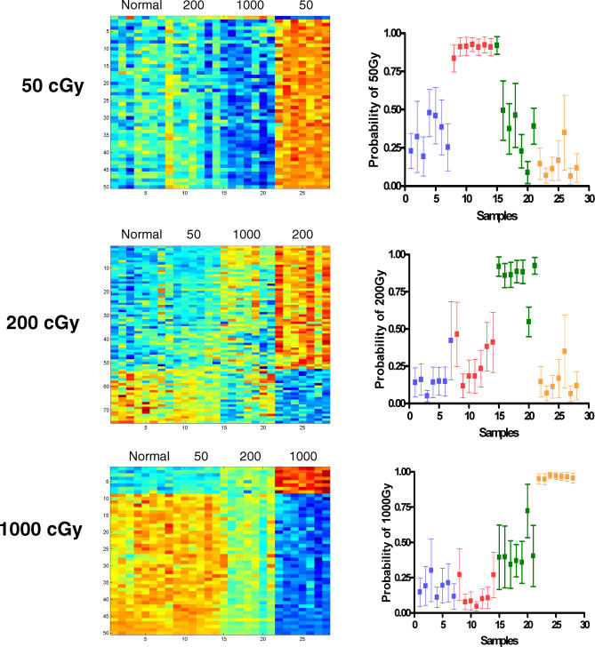
Figure 4. Gene Expression Profiles Can Distinguish Different Levels of Radiation Exposure.
To internally validate the metagene profiles, the predictive capacity of each profile was analyzed with regard to distinguishing nonirradiated samples from each of the irradiated samples. The heatmaps in the left depict the expression profiles of genes selected to discriminate the dose of radiation; high expression is depicted as red, and low expression is depicted as blue; the ranges of relative expression levels are the same as in Figure 3: 0 versus 50 cGy range from 0.29 to 10.5, 0 versus 200 cGy range from 0.26 to 26.3, and 0 versus 1,000 cGy range from 0.17 to 48.77. The right graphs depict a leave-one-out cross validation analysis to demonstrate that in each case, the profiles developed for a particular radiation dose predicted the relevant samples with a high level of accuracy. The samples from normal (nonirradiated) mice are represented as blue, samples from 50-cGy irradiated are red, samples from 200-cGy irradiated are green, and samples from 1,000-cGy irradiated mice are orange. As shown, the predictors for 50-cGy, 200-cGy, and 1,000-cGy irradiation misclassified only one sample out of 28 analyzed in each case, indicating a high level of accuracy of prediction.

