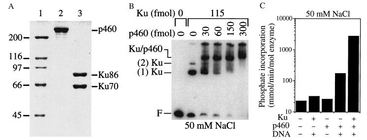
Figure 1.
Analysis of purified p460 and Ku. (A) SDS/PAGE. Purified p460 (lane 2, 350 ng) and Ku (lane 3, 350 ng) were resolved by SDS/PAGE in 8% gels and stained with Coomassie blue. Size markers (lane 1) are defined by their molecular masses in kilodaltons. (B) EMSA. Labeled f32 DNA (0.2 ng) was incubated with Ku (18 ng) and increasing amounts of p460 in 5 μl of buffer B containing 50 mM NaCl and analyzed by EMSA. F denotes the position of free probe. (C) Kinase activity. Peptide substrate (5 μg) was incubated with various combinations of Ku (11 ng), p460 (35 ng), and linear plasmid DNA (300 ng) in 5 μl of buffer C containing 50 mM NaCl, and kinase activity was determined.

